
Clubfoot (CTEV) in Babies: Signs, Diagnosis & Physio Care
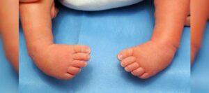
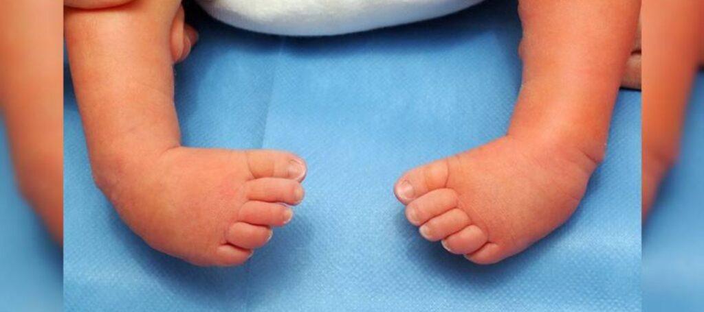
For most new parents in Malaysia, the first newborn check-up is a moment of pure relief, ten fingers, ten toes, all present and accounted for. But for roughly 1 to 2 in every 1,000 babies, that first check reveals something unexpected: a tiny foot that sits turned inward or downward, stiff, and resistant to straightening. This is CTEV, or clubfoot, and it’s one of the most common structural conditions paediatricians pick up at birth.
What Is CTEV? Understanding the Structural Deformity
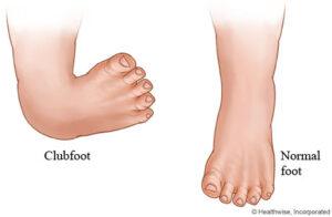
CTEV stands for Congenital Talipes Equinovarus. It’s a structural condition present from birth, where a baby’s foot bones, tendons, and ligaments develop in a fixed, twisted position rather than the typical flat-footed newborn posture. Unlike a foot that simply looks turned because of cramped space in the womb, true CTEV is rigid, it will not passively straighten out, even with gentle hands-on guidance.
CTEV vs. Positional Clubfoot: Know the Difference
Not every “turned-in” newborn foot is CTEV. Many are positional clubfoot, and telling the two apart early changes the entire treatment path.
Feature | Positional Clubfoot | Congenital Clubfoot (CTEV) |
Cause | Cramped foetal positioning in the womb | Structural tendon and bone development |
Flexibility | Soft, corrects with gentle hand pressure | Rigid, resists manual correction |
Typical Course | Resolves within weeks, often with light stretching | Requires formal casting or bracing |
Long-Term Risk if Ignored | Minimal | Abnormal gait, joint strain, mobility issues |
How CTEV Affects a Baby’s Growing Foot
The Biomechanics Behind the Twisted Position
CTEV isn’t a single misalignment it’s a combination of tight, shortened soft tissue structures pulling the foot and ankle joints out of their normal resting position. Because a newborn’s bones are still largely cartilage, this soft-tissue tightness has a powerful effect on how the foot sits and eventually bears weight.
The Four-Part Pattern Breakdown
- Hindfoot Equinus
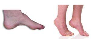
The heel is pulled upward, similar to a tip-toe position, due to a tight Achilles tendon.
- Hindfoot Varus
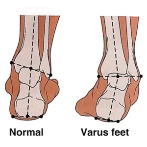
The heel tilts inward under the ankle.
- Forefoot Adduction
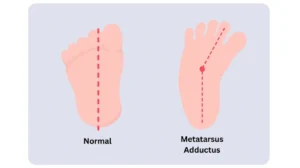
The front of the foot curves inward, toward the midline of the body.
- Talonavicular Subluxation
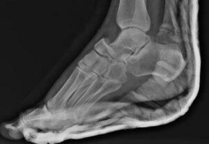
A key bone in the midfoot (the navicular) sits shifted out of its normal alignment with the ankle bone.
Together, these four elements create the classic clubfoot appearance, and explain why the deformity can’t simply be “massaged out” at home.
How Is CTEV Diagnosed?
Clinical Examination
The good news for anxious parents: CTEV is almost always diagnosed through a straightforward physical examination. A paediatric specialist assesses the foot’s flexibility, range of motion, and whether it can be passively guided into a normal position, no invasive testing required.
Prenatal Ultrasound Screening
Some cases are flagged during a routine prenatal scan. However, a prenatal flag isn’t always the final answer, around 10 to 20% of flagged babies turn out to have a normal or mildly positional foot after birth, while a smaller proportion turn out to be more structurally complex. This is exactly why a hands-on postnatal exam remains essential, even after a prenatal scan.
Myth vs. Fact: Common Misconceptions About Clubfoot Treatment
- Myth: Gentle massage or home stretching alone can correct true CTEV.
- Fact: True CTEV requires structural correction — typically serial casting — because the tendons and joint alignment involved cannot be reshaped by soft tissue massage alone.
- Myth: Once the cast comes off, treatment is complete.
- Fact: Removing the cast only corrects the position. Without consistent bracing afterward, relapse is common — bracing is what locks the correction in for good.
Treatment & Recovery: The Ponseti Method and Beyond
Serial Casting (Ponseti Method)
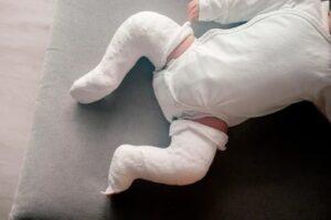
The Ponseti method is the current gold standard for CTEV. It involves weekly, gentle manipulation of the foot followed by casting, gradually repositioning the bones and soft tissue over roughly 4 to 6 weeks.
Tenotomy
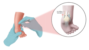
Many babies also need a minor Achilles tenotomy — a quick, low-risk procedure to release residual tightness in the Achilles tendon, usually performed at the final stage of casting.
Bracing & the Role of Physiotherapy in Long-Term Success
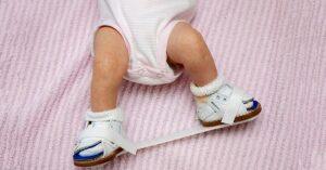
Correction doesn’t end when the cast comes off. A foot abduction brace is worn full-time at first, then part-time, often until age 4 to 5, to hold the corrected position while the child grows. This is where consistent physiotherapy support makes the biggest difference, guiding parents through stretching routines, monitoring gait development as the child starts standing and walking, and catching early signs of relapse before they progress.
Finding Expert Paediatric Physiotherapy Support
Parents searching for paediatric physiotherapy Kuala Lumpur, a trusted child physiotherapist near me, or specialised physiotherapy Puchong services can find experienced, family-focused care at Wai Physio. Beyond CTEV support, our team works with growing children on posture, gait, and developmental milestones, ensuring your child’s foundation for movement is a strong one.
Take Action: Give Your Baby the Best Start
A CTEV diagnosis is not a life sentence, it’s a starting point. With early detection, the right treatment plan, and consistent bracing and physiotherapy support, children with clubfoot grow up walking, running, and playing just like their peers.
Book a paediatric physiotherapy assessment at Wai Physio today and give your baby’s feet the best possible start!